
A. The Heart
1. Has 4 chambers
a. Right Atrium b. Left Atrium
c. Right Ventricle
d. Left Ventricle
2. Made up of cardiac muscle
Heart walls have 3 layers:
a. epicardium
b. myocardium
c. endocardium
3. Myocardium: mass of cells connected by gap junctions
a. this means that an action potential originating anywhere in a myocardium will always be transmitted to all cells of the myocardium
b. these myocardia are separated by a layer of dense connective tissue called the septum
-this will not conduct impulses
4. Has Gap Junctions

a. occur when the cell membranes of adjacent cells are extremely close together b. these membranes appear to share gates through which ions and molecules can pass
from one cell to the next
c. gap junctions are areas of low electrical resistance, meaning that impulses can
easily pass from one cardiac muscle cell to another
d. no gap junctions between atria and ventricles
5. Heart rate
a. autorhythmicity: heart has own internal mechanism that fires on its own
b. Automatic cells have "automatic gates" that open to allow calcium
ions to diffuse into the cells to bring about depolarization of the membrane.
Automatic potassium gates then open allowing potassium to diffuse out to bring about repolarization of the membrane.
d. several areas of the heart are made up of such automatic cells:

2). Atrioventricular (AV) node
3). Atrioventricular (AV) bundle
4). Right and Left Bundle branches
5). Purkinje fibers
e. These cells have different built-in rates. E.g. SA node cells - fastest rate - normally
f. because the SA nodes cells have the fastest rhythm, these cells control the rest of the heart
*** Thus the SA node functions as the pacemaker of the heart ***
g. the impulse that originates at the SA nodes transmits from the atria down into the ventricles
h. Order of stimulation is: (1 - 5 ; from above)
6. Ventricular Myofiber Action Potential

a. chain reaction of depolarization waves (uplimbs of spikes) b. chain reaction of repolarization waves (downlimbs of spikes)
B. Flow of blood through the body
1. systole: contraction
2. diastole: relaxation
3. arteries: carry blood away from the heart
4. veins: carry blood to the heart
5. flow of blood Systemic and Pulmonary Circulation
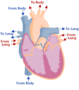
1). superiorly through superior vena cava
2). inferiorly through inferior vena cava b. right atrium contracts pumping deoxygenated blood through the tricuspid valve into the Right Ventricle
c. deoxygenated blood enters the right ventricle
d. right ventricle contracts pumping deoxygenated blood through the pulmonary
valve into the pulmonary trunk then pulmonary arteries
e. deoxygenated blood is transported to the lungs where Carbon Dioxide is
replaced with Oxygen
f. oxygenated blood goes from the lungs through the pulmonary veins into the Left atrium
g. left atrium contracts pumping oxyenated blood through the bicuspid valve into the left ventricle
h. left ventricle contracts pumping oxygenated blood through the aortic valve into the aorta
i. From here the oxygenated blood travels through the arteries to capillaries where gas exchange takes place. Oxygen moves from the capillaries and Carbon Dioxide is picked up. The blood is once again deoxygenated and returns to the right atrium to start process over again.
*** It is important to remember that both atria are contracting at the same time...as are the ventricles***
C. Blood Pressure: the pressure that is exerted by blood against the walls of the blood vessels
1. Systole, contraction, is the higher number
2. Diastole, relaxation, is the lower number
3. Heart sounds
a. LUB - 1st sound heard - caused by closure of cuspid valves
b. DUB - 2nd sound heard - caused by semilunar valves
4. Three important factors affecting blood pressure are:
a. Stroke volume: amount of blood pumped out by ventricles with each contraction
b. Diameter of the vessel: decrease diameter, increase pressure
c. Viscosity of blood: thickness of blood; thinner blood = lower blood pressure
5. Blood pressure is regulated by
a. kidneys
b. Autonomic Nervous system- influences the heart rate and stroke volume
6. Pulse - expansion and contraction of artery produced by waves of pressure caused by
ejection of blood from the left ventricle of the heart as it contracts. Normal is 60-80 bpm.
7. Blood pressure is maintained through activity of the
a. baroreceptors
1) these are stretch receptors (specialized nerve endings)
2) located in the aortic arch and carotid sinuses
3) increase in blood pressure causes the walls of the vessels to stretch thus stimulating the nerves
4) decrease in pressure reduces the stimulation = fewer impulses
5) impulses from the baroreceptors travel to the medulla
b. chemoreceptors
1) measure O2, CO2 content of blood, pH of blood
2) located in the aortic arch and carotid sinuses
3) BP too low, decreases pH, increases CO2, sends signal in order to increases heart rate and vice versa
8. Hypertension - high blood pressure
a. high blood pressure may cause enlargement of heart, heart to work harder, congestive heart failure
b. approx. 20% of all US adults suffer from hypertenstion
c. Normally a systolic pressure over 140 and diastolic pressure over 100.
d. A dangerous condition because high aterial pressure makes it more difficult for the heart to
eject blood causing the heart to work harder leading to congestive heart failure.
High pressure may also damage cerebral blood vessels which may cause strokes.
D. ECGs
1. also called EKGs - stand for electrocardiograms
2. ECGs measure the heart's electrical activity
3. The transmission of electrical impulses through the heart generates a current that travels through the whole body.
4. Thus, electrodes placed on the skin can detect the electrical impulses generated by the heart
5. Graphic representation showing the heart's electrical activity and time interval of that activity can aid those in the medical field
a. to detect arrhythmia - any deviation from normal pattern of heart beat
b. help diagnose damage to the heart from a MI - myocardium infarction
6. Cardiac cycle

b. QRS complex: depolarization and contraction of the ventricles
c. T wave: repolarization of ventricles
d. P-R intervals: length of time in between depolarization of atria and depolarization of ventricles

0 comments:
Post a Comment